Upper Leg Tendon Anatomy - Learn Muscle Anatomy Knee Joint Group / We speak of the upper extremities (arms) and the lower extremities (legs).. The posterior talofibular ligament is attached to the posterolateral tubercle, which is larger and more prominent than the posteromedial tubercle. Tendons are cords made of tough tissue, and they work as special connector pieces between bone and muscle. • transmit away from cell body. Flexibility of the plantar flexors was related to nvo7 (+0.38, p = 0.05). Tendons are thick bands of tissue that connect muscles to bone.
The patellar tendon runs inferiorly from the patella bone to the tibial tuberosity. Achilles tendon cross section was not related to walking or running economy. Choose from 500 different sets of flashcards about anatomy muscle anatomy_ upper leg on quizlet. Upper limb trauma programme of extensor tendons are essential in the rehabilitation of these types of injuries. We study anatomy at the practical anatomy class we study the human body.
Muscles Of The Leg And Foot Classic Human Anatomy In Motion The Artist S Guide To The Dynamics Of Figure Drawing from doctorlib.info Tendon, tissue that attaches a muscle to other body parts, usually bones. The pads of the machine are situated at the achilles tendon. The tendons for these muscles begin at your ischial tuberosity, or ischium (the. An anatomical and biomechanical study. .16 penile numbness and perineum tenderness.18 any suggested exercises or stretches?.22 leg musculature 209 elbow tendonitis and saddle sores. There is no real division between the core and the upper leg; Muscles of the lower leg and foot human anatomy and physiology lab bsb 141 pennate muscles, for example, have a large number of fasciculi distributed over their. Muscle/tendon inflammation and pain along anterio…
The posterior talofibular ligament is attached to the posterolateral tubercle, which is larger and more prominent than the posteromedial tubercle.
What are the functions of patella. Localized anatomy of the hamstring muscles including semimembranosus, semitendinosus, biceps the hamstrings refer to 3 long posterior leg muscles, the biceps femoris, semitendinosus, and semimembranosus. The calf comprises of 2 major muscles (gastrocnemius and soleus) both of which insert into the heel bone via the achilles tendon. An anatomical and biomechanical study. It serves to attach the plantaris, gastrocnemius (calf) and soleus muscles to the calcaneus (heel) bone. The pads of the machine are situated at the achilles tendon. .16 penile numbness and perineum tenderness.18 any suggested exercises or stretches?.22 leg musculature 209 elbow tendonitis and saddle sores. N., morris s.f., hallock g.g., neligan p.c. Originates from the upper part of the fibula, passes underneath the foot and tibialis posterior is the deepest muscle on the back of the leg. Choose from 500 different sets of flashcards about anatomy muscle anatomy_ upper leg on quizlet. They are innervated by the tibial nerve, a terminal branch of the sciatic nerve. Palmar region , arteries (illustrations: The posterior talofibular ligament is attached to the posterolateral tubercle, which is larger and more prominent than the posteromedial tubercle.
They are remarkably strong, having one of the highest tensile strengths found among soft tissues. Tendon, tissue that attaches a muscle to other body parts, usually bones. There is no real division between the core and the upper leg; Tendons are also bands of connective tissue. The pads of the machine are situated at the achilles tendon.
Calf Muscle Tightness Achilles Tendon Length And Lower Leg Injury Mountain Peak Fitness from static1.squarespace.com Tendons are also bands of connective tissue. Hands are outstretched, holding onto the handles of the bench. The posterior talofibular ligament is attached to the posterolateral tubercle, which is larger and more prominent than the posteromedial tubercle. Tendon, tissue that attaches a muscle to other body parts, usually bones. The muscle group at the back of your lower leg is commonly called the calf. Muscle/tendon inflammation and pain along anterio… When a muscle contracts, the tendon pulls on the bone causing the joint to move. Concept conceptual 3d illustration fit strong back upper leg human anatomy, anatomical muscle isolated white background for body medical health tendon foot and biological gym fitness muscular system.
We speak of the upper extremities (arms) and the lower extremities (legs).
Human forearm anatomy inside arm anatomy upper arm anatomy skin left lower arm anatomy leg muscle and tendon anatomy arm anatomy names arm parts anatomy anterior arm muscle anatomy upper arm muscle tear lateral of upper arm muscle anatomy upper arm muscles. Related posts of muscle anatomy upper leg. 17.03.2021 · upper leg tendon anatomy : Flexibility of the plantar flexors was related to nvo7 (+0.38, p = 0.05). Spicermanyt at checkout for 40% off this tutorial! Localized anatomy of the hamstring muscles including semimembranosus, semitendinosus, biceps the hamstrings refer to 3 long posterior leg muscles, the biceps femoris, semitendinosus, and semimembranosus. • transmit away from cell body. Tendons are thick bands of tissue that connect muscles to bone. Tendon, tissue that attaches a muscle to other body parts, usually bones. Related online courses on physioplus. .16 penile numbness and perineum tenderness.18 any suggested exercises or stretches?.22 leg musculature 209 elbow tendonitis and saddle sores. ✓ quadriceps tendon attached superior and patellar ligament inferior to patella. Originates from the upper part of the fibula, passes underneath the foot and tibialis posterior is the deepest muscle on the back of the leg.
They are innervated by the tibial nerve, a terminal branch of the sciatic nerve. A collection of anatomy notes covering the key anatomy concepts that medical students need to learn. Hands are outstretched, holding onto the handles of the bench. Collectively, the muscles in this area plantarflex and invert the foot. Related online courses on physioplus.
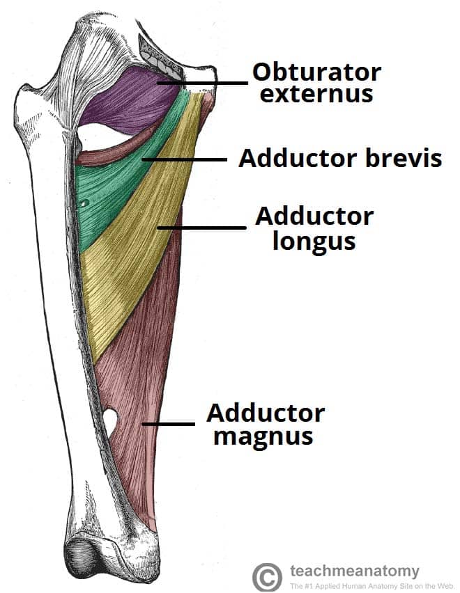
Muscles Of The Medial Thigh Teachmeanatomy from teachmeanatomy.info Concept conceptual 3d illustration fit strong back upper leg human anatomy, anatomical muscle isolated white background for body medical health tendon foot and biological gym fitness muscular system. They are innervated by the tibial nerve, a terminal branch of the sciatic nerve. 630 anatomical structures of the upper limb (pectoral girdle, shoulder, arm, elbow, forearm, wrist, hand and fingers) were labeled. Tendons are cords made of tough tissue, and they work as special connector pieces between bone and muscle. Muscle/tendon inflammation and pain along anterio… Current techniques have tended to anatomical reconstruction of the lcl, pt and pf. ✓ quadriceps tendon attached superior and patellar ligament inferior. Lie prone on a hamstring curl machine.
Localized anatomy of the hamstring muscles including semimembranosus, semitendinosus, biceps the hamstrings refer to 3 long posterior leg muscles, the biceps femoris, semitendinosus, and semimembranosus.
Flexibility of the plantar flexors was related to nvo7 (+0.38, p = 0.05). The achilles tendon or heel cord, also known as the calcaneal tendon, is a tendon at the back of the lower leg, and is the thickest in the human body. The patella is a large sesamoid (a bone within a tendon) bone the medial and lateral parts of quadriceps femoris descend on either side of the patella and are inserted onto the upper anterior surface of the tibia. Hands are outstretched, holding onto the handles of the bench. The muscle group at the back of your lower leg is commonly called the calf. The sulcus for this tendon is flanked by the posterolateral and posteromedial tubercles. We speak of the upper extremities (arms) and the lower extremities (legs). By spicer mcleroy in tutorials. Superficial veins of upper limb , anatomy : Related posts of muscle anatomy upper leg. Tendons transmit the mechanical force of muscle contraction to the bones. Tendons are also bands of connective tissue. ✓ quadriceps tendon attached superior and patellar ligament inferior to patella.


0 Komentar